Afr. J. Parasitol. Mycol. Entomol. , 4(1), 2; doi:10.35995/ajpme04010002
Article
Evaluation of Indoor Fungal Contamination and Cycloheximide Susceptibility of Airborne Molds in Sourô Sanou Teaching Hospital, Burkina Faso—Hospital Air Fungi in Burkina Faso
1
Institut Supérieur des Sciences de la Santé, Université Nazi BONI, 01 BP 1091 Bobo-Dioulasso 01, Burkina Faso
2
Laboratoire de Recherche, Centre MURAZ, 01 BP 390 Bobo-Dioulasso 01, Burkina Faso
3
Unité de Formation et de Recherche en Sciences de la Santé, Université Lédéa Bernard Ouédraogo, 01 BP 346 Ouahigouya 01, Burkina Faso
4
Laboratoire d’Analyses Biomédicales, Centre Hospitalier Universitaire Régional de Ouahigouya, 01 BP 36 Ouahigouya 01, Burkina Faso
5
Laboratoire de Parasitologie-Mycologie, Centre Hospitalier Universitaire Sourô SANOU, 01 BP 676 Bobo-Dioulasso 01, Burkina Faso
*
Corresponding author: cisse_m@yahoo.fr; Tel.: +226-71326769
How to Cite: Cissé, M.; Nakanabo Diallo, S.; Sanou, R.S.; Yerbanga, I.W.; Bamba, S. Evaluation of Indoor Fungal Contamination and Cycloheximide Susceptibility of Airborne Molds in Sourô Sanou Teaching Hospital, Burkina Faso—Hospital Air Fungi in Burkina Faso. Afr. J. Parasitol. Mycol. Entomol. 2026, 4(1): 2; doi:10.35995/ajpme04010002.
Received: 27 December 2025 / Accepted: 21 July 2026 / Published: 20 August 2026
Abstract
:Background: Fungi are common in indoor environments and can cause various diseases. This study aimed to evaluate the extent of fungal contamination in hospital rooms and to assess the susceptibility of mold species isolated from indoor air to cycloheximide. Methods: A prospective cross-sectional study was conducted from November 2023 to February 2024 in five departments of the Sourô Sanou Teaching Hospital, Burkina Faso. Fifty-seven indoor air samples were collected using the passive sedimentation method. The samples were incubated at 30 °C for 24 h, and the resulting colonies were purified. Fungal identification was performed based on macroscopic and microscopic characteristics, and the isolated molds were tested for susceptibility to cycloheximide (0.4 mg/mL). Results: Overall, fungi were recovered from 70.2% (40/57) of air samples, with molds representing the majority of the fungal isolates (99.1%, 224/226). Among the 224 mold isolates identified, A. niger was the most prevalent species (48.7%, 109/224), followed by A. flavus (14.7%, 33/224), Rhizopus spp. (12.5%, 28/224), and A. fumigatus (6.7%, 15/224). The overall cycloheximide inhibition rate was 71.4% (160/224). Lower inhibition rates were observed for A. fumigatus (46.7%), A. versicolor (33.3%), A. flavus (27.3%), Penicillium spp. (27.3%), whereas A. terreus showed complete resistance. Conclusions: These findings underscore the importance of environmental monitoring in preventing and controlling hospital-acquired fungal infections. They also indicate that molds are not uniformly inhibited by cycloheximide, which may influence the accuracy of culture-based diagnostics.
Keywords:
Airborne fungi; hospital environment; mold species; cycloheximide susceptibility; Burkina Faso1. Introduction
Fungal infections have emerged as a major cause of morbidity and mortality in hospitals worldwide. Recent estimates suggest that approximately 6.5 million cases of invasive fungal infections occur each year, resulting in about 3.8 million deaths, of which 2.5 million (68%) are directly attributable to fungal diseases [1]. Aspergillus and Candida are the most common fungal genera responsible for severe infections, accounting for nearly 15% of hospital-acquired infections [2]. Among them, Aspergillus species, particularly A. fumigatus, represent the second leading cause of invasive fungal infections in healthcare settings, with reported mortality rates reaching up to 58% [3]. Another uncommon yet highly fatal infection is mucormycosis, caused by molds belonging to the order Mucorales [4].
The increasing emergence of antifungal-resistant fungi has further complicated the management of invasive fungal infections worldwide. Environmental molds, particularly Aspergillus fumigatus, may harbor intrinsic or acquired resistance to antifungal agents, especially triazoles, partly driven by the widespread use of agricultural azole fungicides and prolonged clinical antifungal exposure [5,6,7]. Antifungal resistance is associated with therapeutic failure, prolonged hospitalization, and increased mortality, especially among immunocompromised patients. In the context of the global antimicrobial resistance crisis, surveillance of environmental fungal reservoirs and their susceptibility profiles has become an important component of infection prevention and antimicrobial stewardship strategies [6,8].
The primary route of transmission of invasive fungal infections in susceptible individuals is exposure to mold spores, which are ubiquitous in soil, in air, and on surfaces [9]. These spores readily become airborne and can disseminate throughout hospital environments, where numerous mold genera, including Alternaria, Cladosporium, Aspergillus, Penicillium, Rhizopus, Fusarium, and Mucor, have been detected [3,4,10,11,12,13]. A recent systematic review of airborne fungal monitoring in healthcare facilities further identified Aspergillus, Penicillium, Alternaria, Cladosporium, and Rhizopus as the most frequently recovered genera and emphasized the absence of internationally standardized protocols and guidelines for fungal surveillance in hospital environments [14]. Assessing their presence and diversity in healthcare facilities is therefore essential, particularly in wards caring for immunocompromised or otherwise vulnerable patients [3,4].
Molds are frequently encountered in microbiology laboratories and are generally considered environmental contaminants [15]. To prevent contamination of fungal cultures, Sabouraud dextrose agar is commonly supplemented with cycloheximide. This compound inhibits eukaryotic protein synthesis, including that of microscopic fungi (eumycetes), by blocking both the initiation and elongation phases of protein synthesis translation [16]. However, data on the antifungal activity of cycloheximide remain limited. Furthermore, the existing studies are outdated, and contemporary evidence regarding its efficacy and use in sub-Saharan Africa is scarce [17,18,19,20].
In Burkina Faso, information on the fungal component of indoor air quality in hospital settings remains limited. The few available studies are outdated and were conducted exclusively at Sourô Sanou Teaching Hospital in Bobo-Dioulasso, the country’s second-largest city. Reported airborne fungal contamination rates ranged from 66.9% to 85.5%, with Aspergillus species identified as the predominant isolates [21,22,23]. The antifungal activity of cycloheximide, however, has not been investigated against these environmental fungi. This study was therefore undertaken to characterize the antifungal activity of cycloheximide on airborne fungal isolates collected from hospital indoor environments in Burkina Faso, thereby addressing an important knowledge gap.
2. Materials and Methods
2.1. Study Design and Study Site
This prospective cross-sectional study was conducted during the dry season, from November 2023 to February 2024 at the Sourô Sanou Teaching Hospital in Bobo-Dioulasso, the second-largest city in Burkina Faso. The hospital has a capacity of 550 beds distributed across six departments and serves as the country’s second major referral center.
2.2. Sampling
Indoor air sampling was performed in five departments selected because they represent areas at increased risk of exposure to airborne fungal pathogens: the medical emergency, pediatric emergency, surgical emergency, intensive care, and laboratory departments. These departments were prioritized because they manage critically ill or vulnerable patients and/or perform microbiological activities in which monitoring airborne fungal contamination is particularly relevant. Samples were collected randomly within each department using the passive sedimentation technique with 90 mm Petri dishes (CITOTEST Scientific Co., Ltd., Nanjing, China) containing Sabouraud dextrose agar. A total of 57 air samples were collected, comprising 14 samples from the laboratory department, 14 from the medical emergency department, 11 from the intensive care unit, 10 from the pediatric emergency department, and 8 from the surgical emergency department. The placement of the Petri dishes was standardized across all sampled departments. In each department, plates were positioned at comparable predetermined locations, including beneath air conditioners, adjacent to windows, and on shelves whenever these features were available. When the physical configuration of a department differed, the closest equivalent locations were selected to ensure consistency in the sampling procedure. All plates were exposed for 24 h before being transported to the Parasitology-Mycology Laboratory for processing.
2.3. Fungi Identification
Samples were incubated at 30 °C for 24 h to detect fungal growth. Positive cultures were subcultured onto Sabouraud dextrose agar supplemented with chloramphenicol and incubated at 30 °C for an additional 24 h for purification as previously described by Yerbanga et al. [23].
Macroscopic identification was based on colony morphology, including color and texture. For microscopic examination, a tape mount preparation was performed by pressing transparent adhesive tape onto the colony and placing it on a drop of lactophenol cotton blue on a glass slide. Fungal structures were examined under a light microscope at 10× and 40× magnification, focusing on the morphology of conidia, hyphae, and reproductive elements, following the procedures described by Yerbanga et al. [23].
2.4. Cycloheximide Susceptibility Testing
The inhibitory activity of cycloheximide against airborne mold fungi was evaluated by subculturing selected isolates onto Sabouraud dextrose agar supplemented with chloramphenicol and cycloheximide (Condalab, Madrid, Spain), containing cycloheximide at a final concentration of 0.4 mg/mL. The dehydrated medium was prepared according to the manufacturer’s instructions by suspending 65.5 g of powder in 1 L of distilled water. The suspension was heated with frequent agitation until completely dissolved, sterilized by autoclaving at 121 °C for 15 min, cooled to 45–50 °C, and aseptically dispensed into sterile 90 mm polystyrene Petri dishes (CITOTEST Scientific Co., Ltd., Nanjing, China). After solidification, the plates were stored at 2–8 °C until use. Selected mold isolates were inoculated onto the prepared medium and incubated at 30 °C for 48 h. Following incubation, cultures were examined for visible fungal growth. Isolates showing no growth were considered susceptible to cycloheximide, whereas those exhibiting any visible growth were classified as resistant.
2.5. Data Analysis
All data were entered into Microsoft Excel 2016 and analyzed using SPSS software, version 25 (IBM Corp., Armonk, NY, USA). Descriptive statistical methods were employed to summarize the results as frequencies and proportions.
3. Results
3.1. Identification of Isolated Fungi
Of the 57 air samples collected from the five hospital departments, 40 yielded fungal growth, corresponding to an overall prevalence of airborne fungal contamination of 70.2%.
A total of 226 fungal isolates were identified, of which molds accounted for 99.1% (224/226). Aspergillus was the predominant genus (75.2%; 170/226), followed by Rhizopus (12.4%; 28/226) and Penicillium (4.0%; 9/226). Among yeasts, only the Candida genus was detected, representing 0.9% (2/226) of all isolates (Figure 1). Within the mold isolates, A. niger was the most frequent species (48.7%; 109/224), followed by A. flavus (14.7%; 33/224), Rhizopus spp. (12.5%; 28/224), and A. fumigatus (6.7%; 15/224) (Table 1).
Figure 1.
Prevalence of fungal genera detected in air samples at the Sourô Sanou Teaching Hospital.
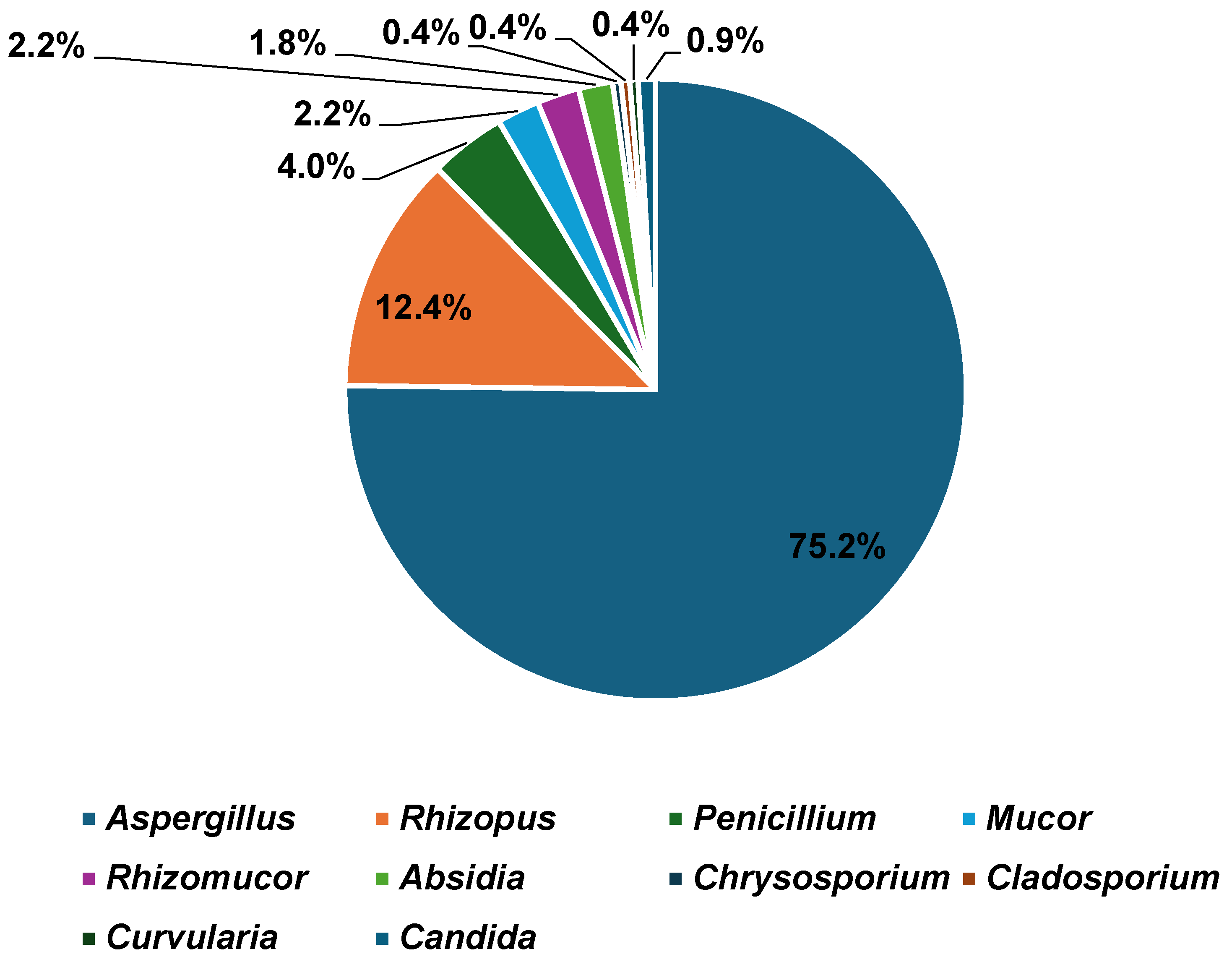

Table 1.
Distribution of the proportion of different mold species identified (n = 224).
| Mold Species | Frequency | % |
|---|---|---|
| Aspergillus niger | 109 | 48.7 |
| Aspergillus flavus | 33 | 14.7 |
| Rhizopus spp. | 28 | 12.5 |
| Aspergillus fumigatus | 15 | 6.7 |
| Aspergillus versicolor | 9 | 4.0 |
| Penicillium spp. | 9 | 4.0 |
| Mucor spp. | 5 | 2.2 |
| Rhizomucor miehei | 5 | 2.2 |
| Absidia corymbifera | 4 | 1.8 |
| Aspergillus spp. | 2 | 0.9 |
| Aspergillus terreus | 1 | 0.4 |
| Aspergillus candidus | 1 | 0.4 |
| Chrysosporium keratinophilum | 1 | 0.4 |
| Cladosporium spp. | 1 | 0.4 |
| Curvularia spp. | 1 | 0.4 |
| Total | 224 | 100 |
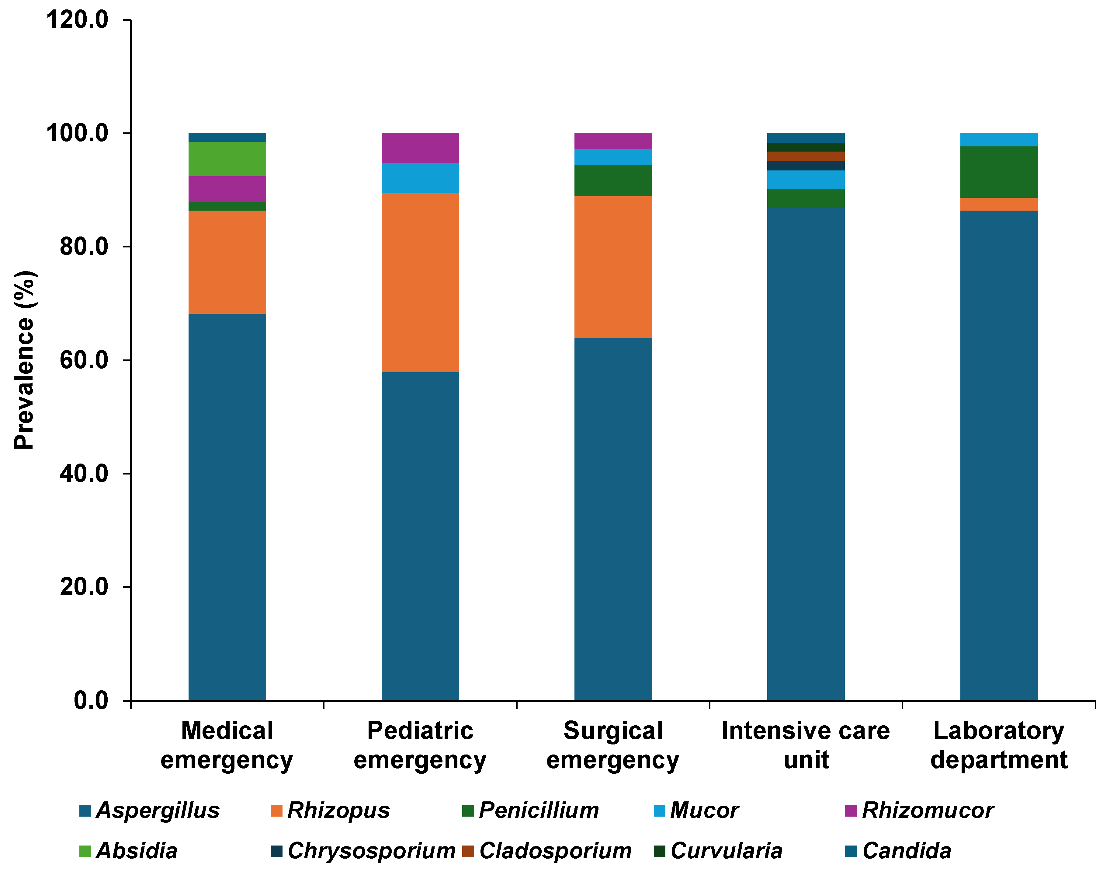
The medical emergency department yielded the highest proportion of fungal isolates (29.2%; 66/226), followed by the intensive care unit (27.0%; 61/226), the laboratory department (19.5%; 44/226), the surgical emergency department (15.9%; 36/226), and the pediatric emergency department (8.4%; 19/226). The distribution of fungal genera and species across departments is presented in Table 2. Aspergillus was the predominant genus in all departments, accounting for 57.9% to 83.6% of isolates. A. niger was the most frequently recovered species in the laboratory, medical emergency, intensive care, and surgical emergency departments, whereas Rhizopus spp. predominated in the pediatric emergency department (31.6%; 6/19). Penicillium spp. were isolated from four of the five departments, while Mucor spp. were detected in all departments except the medical emergency unit. Less frequently recovered fungi, including Absidia corymbifera, Rhizomucor miehei, Chrysosporium sp., Cladosporium sp., Curvularia sp., and Candida spp., were restricted to one or two departments (Figure 2; Table 2).
Table 2.
Distribution of airborne fungal isolates by genus/species and hospital department.
| Fungal Genus/Species | Laboratory (44 Isolates) | Medical Emergency (66 Isolates) | Intensive Care (61 Isolates) | Pediatric Emergency (19 Isolates) | Surgical Emergency (36 Isolates) | |||||
|---|---|---|---|---|---|---|---|---|---|---|
| n | (%) | n | (%) | n | (%) | n | (%) | n | (%) | |
| Aspergillus niger | 28 | 63.6 | 23 | 34.8 | 38 | 62.3 | 3 | 15.8 | 17 | 47.2 |
| Aspergillus flavus | 9 | 20.4 | 11 | 16.7 | 8 | 13.1 | 3 | 15.8 | 2 | 5.6 |
| Aspergillus fumigatus | 1 | 2.3 | 4 | 6.1 | 1 | 1.6 | 5 | 26.3 | 4 | 11.1 |
| Aspergillus versicolor | – | 7 | 10.6 | 2 | 3.3 | – | – | |||
| Aspergillus terreus | – | – | 1 | 1.6 | – | – | ||||
| Aspergillus candidus | – | – | 1 | 1.6 | – | – | ||||
| Aspergillus spp. | – | – | 2 | 3.3 | – | – | ||||
| Rhizopus spp. | 1 | 2.3 | 12 | 18.2 | – | 6 | 31.6 | 9 | 25.0 | |
| Mucor spp. | 1 | 2.3 | – | 2 | 3.3 | 1 | 5.3 | 1 | 2.8 | |
| Rhizomucor miehei | – | 3 | 4.5 | – | 1 | 5.3 | 1 | 2.8 | ||
| Absidia corymbifera | – | 4 | 6.1 | – | – | – | ||||
| Penicillium spp. | 4 | 9.1 | 1 | 1.5 | 2 | 3.3 | – | 2 | 5.6 | |
| Chrysosporium spp. | – | – | 1 | 1.6 | – | – | ||||
| Cladosporium spp. | – | – | 1 | 1.6 | – | – | ||||
| Curvularia spp. | – | – | 1 | 1.6 | – | – | ||||
| Candida spp. * | – | 1 | 1.5 | 1 | 1.6 | – | – | |||
| Total isolates | 44 | 100.0 | 66 | 100.0 | 61 | 100.0 | 19 | 100.0 | 36 | 100.0 |
* Candida isolates were identified only to the genus level using conventional macroscopic and microscopic methods. Species-level identification was not performed. Air samples collected: Laboratory (14), Medical Emergency (14), Intensive Care (11), Pediatric Emergency (10), and Surgical Emergency (8).
Figure 2.
Prevalence of fungal genera detected in air samples at five departments of the Sourô Sanou Teaching Hospital.
Figure 2.
Prevalence of fungal genera detected in air samples at five departments of the Sourô Sanou Teaching Hospital.
3.2. Susceptibility of Mold Species to Cycloheximide
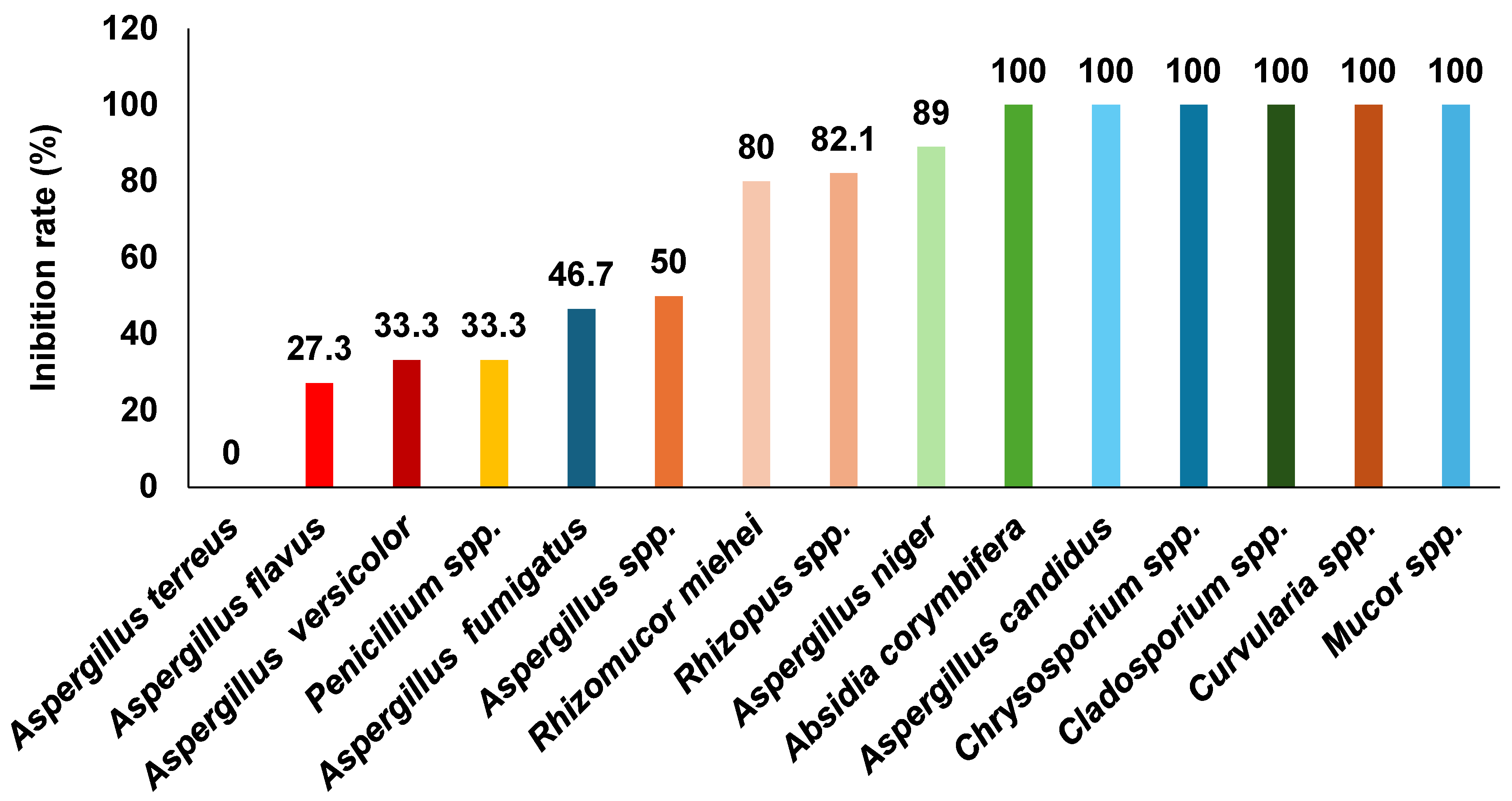
The overall cycloheximide inhibition rate among the mold isolates was 71.4% (160/224). Complete inhibition (100%) was observed for Absidia corymbifera (4/4), Aspergillus candidus (1/1), Chrysosporium spp. (1/1), Cladosporium spp. (1/1), Curvularia spp. (1/1), and Mucor spp. (5/5) (Figure 3). In contrast, lower inhibition rates were recorded for A. versicolor (33.3%; 3/9), A. flavus (27.3%; 9/33), Penicillium spp. (27.3%; 3/11), and A. fumigatus (46.7%; 7/15). Notably, the A. terreus isolate (1/1) exhibited complete resistance to cycloheximide.
Figure 3.
Susceptibility of mold species to cycloheximide.
4. Discussion
The overall prevalence of airborne fungal flora observed in this study (70.2%) was lower than the rates previously reported at the Sourô Sanou Teaching Hospital, which ranged from 83.5% to 88% in 2017 for the central operating room and intensive care unit [21,22], and 86.1% in 2019 for the medical emergency and neonatology units [23]. This difference may be attributed to the smaller number of samples analyzed in the present study (57 versus >200 in earlier investigations) and to temporal variations in environmental conditions influencing airborne fungal load.
Airborne fungal contamination rates reported in hospital environments worldwide vary widely, from 35% to 100% [13,24,25,26,27]. Such variability may reflect differences in climatic conditions, geographical location, hospital infrastructure, ventilation systems, occupancy, and infection prevention practices. A recent systematic review also highlighted substantial heterogeneity in airborne fungal monitoring methods, including differences in sampling techniques, culture media, incubation conditions, and interpretation criteria, emphasizing the need for standardized surveillance protocols in healthcare settings [14].
Because air sampling was conducted exclusively during the dry season (November 2023 to February 2024), the fungal distribution observed in this study should be interpreted within this seasonal context. Climatic factors such as temperature, humidity, rainfall, and wind are known to influence fungal growth, sporulation, and airborne spore dispersion. Consequently, seasonal fluctuations in airborne fungal contamination have been reported in healthcare environments [14]. Longitudinal studies covering both the dry and rainy seasons would provide a more comprehensive understanding of seasonal variations in indoor fungal communities in Burkina Faso.
The predominance of molds observed in this study is consistent with findings from previous investigations in the same hospital [21,22,23] and in other regions [11,12,13,24,25,27,28,29,30]. Molds are ubiquitous, filamentous fungi that produce numerous lightweight spores capable of dispersing through air currents and by human or animal carriers [9,10]. These characteristics facilitate their persistence and rapid colonization of indoor environments, including healthcare facilities.
Aspergillus was the predominant genus in all hospital departments, accounting for 57.9% to 86.9% of recovered isolates. Although Aspergillus predominated across all departments, the relative abundance of other fungal genera varied. Rhizopus spp., for example, were proportionally more frequent in the pediatric emergency (31.6%) and surgical emergency (25.0%) departments, whereas Penicillium spp. were recovered from four of the five departments. These differences may reflect variations in patient turnover, ventilation, environmental conditions, cleaning practices, and the specific activities performed within each department. Similar spatial heterogeneity of airborne fungal communities has been reported in hospital environments elsewhere [14].
The predominance of the Aspergillus genus observed here aligns with reports from Burkina Faso [21,22,23] and other regions [11,12,13,29]. Aspergillus species are cosmopolitan fungi capable of surviving under diverse environmental conditions. Their spores originate from multiple reservoirs, including soil, organic matter, dust, and ventilation systems, and can remain airborne for prolonged periods [9,31]. Their ecological adaptability and efficient spore dispersal likely explain their predominance in the present study.
In contrast, Cladosporium and Penicillium were the predominant genera in studies from Senegal and Portugal, respectively [27,30]. Such regional variation in fungal communities reflects differences in local climate, architecture, and ventilation, as well as variable standards of environmental control [9,27,30,31].
Among the Aspergillus isolates, A. niger was the most frequently identified species (48.7%), consistent with previous findings in Burkina Faso [23] and Tunisia [25]. A. niger is a common environmental contaminant found in food, soil, and indoor air owing to its ability to colonize diverse substrates [9,31]. Although widely distributed, A. niger is less pathogenic to humans than other Aspergillus species [32], typically affecting tissues compromised by infection, trauma, or cerumen accumulation [31]. Notably, A. fumigatus predominated in the same hospital in 2017 [21,22], suggesting temporal shifts in the composition of airborne fungal communities. Similarly, A. fumigatus was predominant in studies from Brazil [12] and Italy [29], whereas Penicillium spp. predominated in Iran [28] and Portugal [30]. These discrepancies may result from environmental and geographic differences, as well as variations in hospital infrastructure and infection control measures.
The overall inhibition rate of cycloheximide in this study (71.4%) was higher than that reported by Fuente et al. in the United Kingdom (40%) [19], likely due to the higher concentration used here (0.4 mg/mL vs. 0.1 mg/mL) and differences in fungal species composition. Conversely, the inhibition rate was lower than that reported in the United States (US) (93.3%) [18]. This may be explained by methodological differences, including a smaller sample size (15 isolates vs. 224), a slightly higher cycloheximide concentration (0.5 mg/mL), and the specific fungal species tested. In the U.S. study, Aspergillus, Mucor, Penicillium, and Rhizopus isolates were all inhibited, whereas only one of the two Cladosporium isolates showed susceptibility [18]. In the present study, inhibition rates were 69.6% against Aspergillus, 100% against Mucor, 33.3% against Penicillium, and 82.1% against Rhizopus.
A complete inhibition rate (100%) was also reported in the United States in 1950 for Aspergillus, Cladosporium, Penicillium, and Rhizopus [17], possibly reflecting the smaller number of isolates tested. Similarly, cycloheximide demonstrated complete inhibitory activity against Absidia, Chrysosporium, Cladosporium, Curvularia, and Mucor isolates in this study, consistent with findings from Chabasse et al. in France [20].
Although two Candida isolates were recovered in the present study, they were identified only to the genus level. Therefore, their response to cycloheximide could not be interpreted at the species level. This is relevant because cycloheximide susceptibility varies considerably among Candida species: some exhibit intrinsic tolerance, whereas others are inhibited at concentrations commonly used in selective culture media, as reported by Dal Pizzol et al. [33]. Species-level identification would therefore be necessary to interpret the cycloheximide growth response of Candida isolates accurately.
This study has several limitations. First, the passive sedimentation method, although simple and cost-effective, has limited sensitivity in highly controlled environments and lacks standardized international reference values, limiting comparisons across studies [34]. Second, environmental assessment was restricted to air sampling; hospital surfaces, medical materials, and reusable medical instruments were not sampled, providing only a partial characterization of the hospital fungal environment [34]. Third, fungal identification relied solely on conventional macroscopic and microscopic methods, which have limited discriminatory power for closely related fungal species and do not allow reliable species-level identification of yeasts such as Candida. Consequently, yeast isolates were identified only to the genus level, and some filamentous fungi may have been misidentified. Fourth, air sampling was not standardized with respect to routine cleaning and disinfection schedules, precluding differentiation between residual contamination and environmental recontamination. Finally, the effect of cycloheximide was evaluated using a single concentration (0.4 mg/mL) from a single manufacturer, limiting the generalizability of the findings. Future studies should integrate air, surface, material, and instrument sampling, employ advanced fungal identification techniques (e.g., chromogenic media, MALDI-TOF mass spectrometry, automated identification systems such as Vitek 2, and molecular methods), standardize sampling in relation to cleaning procedures, and evaluate multiple cycloheximide concentrations.
5. Conclusions
Airborne fungal contamination was frequent in the surveyed hospital departments, with Aspergillus as the predominant genus and A. niger as the most common species. Cycloheximide did not inhibit all environmental molds, particularly A. flavus, A. versicolor, A. fumigatus, A. terreus, and Penicillium spp., underscoring variable susceptibility among isolates. These findings highlight the limitations of cycloheximide-based media for selective inhibition of saprophytic fungi.
Improving fungal diagnostics requires the use of multiple culture media, enhanced environmental monitoring, and efficient air filtration to minimize contamination. Future research should evaluate cycloheximide efficacy across different concentrations and formulations, apply molecular methods for precise species identification, and explore links between airborne fungal load, seasonal variation, and hospital-acquired infections.
Author Contributions
M.C. analyzed the data, wrote the original draft, and participated in the review and editing of the manuscript. R.S.S. performed field data collection and laboratory work under the supervision of S.N.D. S.N.D. supervised field data collection and laboratory work and contributed to data analysis, manuscript review, and editing. I.W.Y. contributed to manuscript review and editing. S.B. conceived the study, designed the methodology, and supervised the study. All authors have read and agreed to the published version of the manuscript.
Funding
No specific funding was received for this study.
Acknowledgments
The authors are grateful to the director and health professionals of the Sourô Sanou Teaching Hospital.
Conflicts of Interest
The authors declare that there are no conflicts of interest.
References
- Denning, D.W. Global incidence and mortality of severe fungal disease. Lancet Infect. Dis. 2024, 24, e428–e438. [Google Scholar] [CrossRef]
- Jones, C.L. The potential environmental role of fungi as a complication in COVID-19 infections. J. Bacteriol. Mycol. Open Access 2020, 8, 6–13. [Google Scholar] [CrossRef]
- Park, J.H.; Ryu, S.H.; Lee, J.Y.; Kim, H.J.; Kwak, S.H.; Jung, J.; Lee, J.; Sung, H.; Kim, S.-H. Airborne fungal spores and invasive aspergillosis in hematologic units in a tertiary hospital during construction: A prospective cohort study. Antimicrob. Resist. Infect. Control 2019, 8, 88. [Google Scholar] [CrossRef] [PubMed]
- Jordan, A.; James, A.E.; Gold, J.A.W.; Wu, K.; Glowicz, J.; Wolfe, F.; Vyas, K.; Litvintseva, A.; Gade, L.; Liverett, H.; et al. Investigation of a Prolonged and Large Outbreak of Healthcare-Associated Mucormycosis Cases in an Acute Care Hospital-Arkansas, June 2019–May 2021. Open Forum Infect. Dis. 2022, 9, ofac510. [Google Scholar] [CrossRef] [PubMed]
- Verweij, P.E.; Chowdhary, A.; Melchers, W.J.G.; Meis, J.F. Azole resistance in Aspergillus fumigatus: Can we retain the clinical use of mold-active antifungal azoles? Clin. Infect. Dis. 2016, 62, 362–368. [Google Scholar] [CrossRef] [PubMed]
- Fisher, M.C.; Alastruey-Izquierdo, A.; Berman, J.; Bicanic, T.; Bignell, E.M.; Bowyer, P.; Bromley, M.; Brüggemann, R.; Garber, G.; Cornely, O.A.; et al. Tackling the emerging threat of antifungal resistance to human health. Nat. Rev. Microbiol. 2022, 62, 362–368. [Google Scholar] [CrossRef] [PubMed]
- Yerbanga, I.W.; Lagrou, K.; Merckx, R.; Diallo, S.N.; Gangneux, J.; Delabarre, A.; Denis, O.; Rodriguez-Villalobos, H.; Montesinos, I.; Bamba, S. First detection of triazole-resistant Aspergillus fumigatus harbouring the TR34/L98H Cyp51A mutation in Burkina Faso. Mycoses 2024, 67, e13732. [Google Scholar] [CrossRef] [PubMed]
- WHO. WHO Fungal Priority Pathogens List to Guide Research, Development and Public Health Action; World Health Organization: Geneva, Switzerland, 2022. [Google Scholar]
- ANOFEL. Parasitoses et Mycoses des Régions Tempérées et Tropicales, 7th ed.; Masson: Paris, France; Elsevier: Amsterdam, The Netherlands, 2021; 998p. [Google Scholar]
- Mareković, I. What’s new in prevention of invasive fungal diseases during hospital construction and renovation work: An overview. J. Fungi 2023, 9, 151. [Google Scholar] [CrossRef] [PubMed]
- Teles de Oliveira, M.; Kelly, N.; Batista, R.; de Souza Gil, E.; Silva Mdo, R.R.; Costa, C.R.; Bara, M.T.F.; Torres, I.M.S. Risks associated with pathogenic fungi isolated from surgical centers, intensive care units, and materials sterilization center in hospitals. Risks associated with pathogenic fungi isolated from critical hospital areas. Med. Mycol. 2020, 58, 881–886. [Google Scholar] [CrossRef] [PubMed]
- Lemos, M.S.C.d.; Higa Junior, M.G.; Paniago, A.M.M.; Melhem, M.d.S.C.; Takahashi, J.P.F.; Fava, W.S.; Venancio, F.A.; Martins, N.M.; Chang, M.R. Aspergillus in the Indoor Air of Critical Areas of a Tertiary Hospital in Brazil. J. Fungi 2024, 10, 538. [Google Scholar] [CrossRef] [PubMed]
- Ghodsi, S.; Nikaeen, M.; Aboutalebian, S.; Mohammadi, R.; Mirhendi, H. Prevalence of fungi and their antifungal and disinfectant resistance in hospital environments: Insights into combating nosocomial mycoses. Antimicrob. Resist. Infect. Control 2025, 14, 37. [Google Scholar] [CrossRef] [PubMed]
- Surwill, D.L.; Cruz, P.; Buttner, M.P.; Pharr, J.R.; Lough, N.; Roehr, T.T. Airborne fungal monitoring in healthcare environments: A systematic review. J. Fungi 2026, 12, 336. [Google Scholar] [CrossRef] [PubMed]
- Blaize, M.; Normand, A.C.; Fekkar, A.; Piarroux, R. Identification des moisissures au laboratoire de routine hospitalière. Rev. Fr. Lab. 2021, 2021, 58–65. [Google Scholar] [CrossRef]
- Sabouraud, R. Maladies du Cuir Chevelu. Les Maladies Cryptogamiques: Les Teignes; Masson et Cie: Paris, France, 1902. [Google Scholar]
- Phillips, G.; Hanel, E. Control of mold contaminants on solid media by the use of actidione. J. Bacteriol. 1950, 60, 104–105. [Google Scholar] [CrossRef] [PubMed]
- Georg, L.; Ajello, L.; Gordon, M. A selective medium for the isolation of Coccidioides immitis. Science 1951, 114, 387–389. [Google Scholar] [CrossRef] [PubMed]
- Fuentes, C.A.; Trespalacios, F.; Baquero, G.F.; Aboulafia, R. Effect of actidione on mold contaminants and on human pathogens. Mycologia 1952, 44, 170–175. [Google Scholar] [CrossRef]
- Chabasse, D.; Contet-Audonneau, N.; Bouchara, J.P.; Basile, A.M. Moisissures, Dermatophytes, Levures. Du Prélèvement au Diagnostic; Biomérieux: Marcy-l’Étoile, France, 2008; 190p. [Google Scholar]
- Segda, G.R. Analyse de la Composition Qualitative de la Flore Fongique de L’environnement du Bloc Opératoire Central du Centre Hospitalier Universitaire Sourô Sanou de Bobo-Dioulasso (Burkina Faso); UFR/SDS, Université Joseph-Ki Zerbo: Ouagadougou, Burkina Faso, 2017. [Google Scholar]
- Matotou, H.R.S.; Sangare, I.; Bisseye, C.; Bouyou Akotet, M.K.; Bamba, S. Biodiversité de la flore fongique isolée au service de réanimation du Centre Hospitalo-Universitaire Souro Sanou de Bobo-Dioulasso, Burkina Faso. Pan Afr. Med. J. 2021, 38, 299. [Google Scholar] [CrossRef] [PubMed]
- Yerbanga, I.W.; Kaboré, R.W.V.; Nana, D.; Diallo-Nakanabo, S.; Rouamba, T.; Gnoumou, N.; Denis, O.; Rodriguez-Villalobos, H.; Montesinos, I.; Bamba, S. A Qualitative assessment of fungal bioaerosols in the indoor air of the Sourô Sanou University Hospital center of Bobo-Dioulasso, Burkina Faso. Afr. J. Parasitol. Mycol. Entomol. 2023, 1, 1. [Google Scholar] [CrossRef]
- D’Alessandro, D.; Cerquetani, F.; Deriu, M.; Montagna, M.; Mura, I.; Napoli, C.; Vescia, N. Evaluation of fungal contamination in operating rooms using a dusting cloth pad: Comparison among different sampling methods. Am. J. Infect. Control 2013, 41, 658–660. [Google Scholar] [CrossRef] [PubMed]
- Neji, S.; Ayadi, A.; Aloulou, M.; Trabelsi, H.; Sellami, H.; Cheikhrouhou, F.; Triki, Z.; Guidara, R.; Makni, F.; Karoui, A.; et al. Etude de la flore fongique des blocs opératoires de l’hôpital de Sfax. Rev. D’hygiène 2014, XXII, 39–42. [Google Scholar]
- Ashuro, Z.; Diriba, K.; Afework, A.; Washo, G.H.; Areba, A.S.; Kanno, G.G.; Hareru, H.E.; Kaso, A.W.; Tesfu, M. Assessment of microbiological quality of indoor air at different hospital sites of Dilla University: A cross-sectional study. Enviorn. Health Insights 2022, 16, 11786302221100048. [Google Scholar] [CrossRef] [PubMed]
- Diongue, K.; Badiane, A.; Seck, M.; Ndiaye, M.; Diallo, M.; Diallo, S.; Sy, O.; Ndiaye, J.; Faye, B.; Ndir, O.; et al. Composition qualitative de la flore fongique de l’environnement de 07 services à risque d’infections fongiques au CHU Aristide Le Dantec (Dakar) Qualitative fungal composition of services at risk of nosocomial infections at Aristide Le Dantec Hospital. J. Mycol. Med. 2015, 25, e39–43. [Google Scholar] [CrossRef] [PubMed]
- Azimi, F.; Naddafi, K.; Nabizadeh, R.; Hassanvand, M.S.; Alimohammadi, M.; Afhami, S.; Musavi, S.N. Fungal air quality in hospital rooms: A case study in Tehran, Iran. J. Environ. Health Sci. Eng. 2013, 11, 30. [Google Scholar] [CrossRef] [PubMed]
- Caggiano, G.; Napoli, C.; Coretti, C.; Lovero, G.; Scarafile, G.; De Giglio, O.; Montagna, M.T. Mold contamination in a controlled hospital environment: A 3-year surveillance in southern Italy. BMC Infect. Dis. 2014, 14, 595. [Google Scholar] [CrossRef] [PubMed]
- Cabo Verde, S.; Almeida, S.M.; Matos, J.; Guerreiro, D.; Meneses, M.; Faria, T.; Botelho, D.; Santos, M.; Viegas, C. Microbiological assessment of indoor air quality at different hospital sites. Res. Microbiol. 2015, 166, 557–563. [Google Scholar] [CrossRef] [PubMed]
- Egbuta, M.A.; Mwanza, M.; Babalola, O.O. Health risks associated with exposure to filamentous fungi. Int. J. Environ. Res. Public Health 2017, 14, 719. [Google Scholar] [CrossRef] [PubMed]
- Person, A.K.; Chudgar, S.M.; Norton, B.L.; Tong, B.C.; Stout, J.E. Aspergillus niger: An unusual cause of invasive pulmonary aspergillosis. J. Med. Microbiol. 2010, 59, 834–838. [Google Scholar] [CrossRef] [PubMed]
- Dal Pizzol, M.; Freitas, E.C.; Locatelli, C.; Guareze, F.; Reginatto, P.; Machado, G.; Fuentefria, A.; Marinho, D. Antifungal efficacy and safety of cycloheximide as a supplement in optisol-GS. Drug Des. Dev. Ther. 2021, 15, 2091–2098. [Google Scholar] [CrossRef] [PubMed]
- Tomazin, R.; Matos, T. Mycological Methods for Routine Air Sampling and Interpretation of Results in Operating Theaters. Diagnostics 2024, 14, 288. [Google Scholar] [CrossRef] [PubMed]
© 2026 Copyright by Authors. Licensed as an open access article using a CC BY 4.0 license.
