
Afr. J. Parasitol. Mycol. Entomol. , 3(2), 14; doi:10.35995/ajpme03020014
Article
Diagnostic Performance of the Sysmex XN-31™ Automated Analyzer for Detecting Asymptomatic Malaria Among Blood Donors: A Comparative Cross-Sectional Study at the Regional Blood Transfusion Center of Abidjan, Côte d’Ivoire
Pulchérie Christiane Kiki Barro 1,2,*,
Eugénie Yaudé 1,
Kpongbo Etienne Angora 2 ,
Lemaitre Hardy Emmanuella Kouadio 2,
Assohoun Jean Sébastien Miézan 1,2,
Valérie Akoua Bédia-Tanoh 2,
Gnanyo Massafoma Estelle Koné 2,
Abo Henriette Vanga-Bosson 2,
Fulgence Kondo Kassi 2,
Abibatou Konaté-Touré 2,
Vincent Djohan 2,
Eby Ignace Hervé Menan 2
and
William Yavo 1,2
,
Lemaitre Hardy Emmanuella Kouadio 2,
Assohoun Jean Sébastien Miézan 1,2,
Valérie Akoua Bédia-Tanoh 2,
Gnanyo Massafoma Estelle Koné 2,
Abo Henriette Vanga-Bosson 2,
Fulgence Kondo Kassi 2,
Abibatou Konaté-Touré 2,
Vincent Djohan 2,
Eby Ignace Hervé Menan 2
and
William Yavo 1,2
,
Lemaitre Hardy Emmanuella Kouadio 2,
Assohoun Jean Sébastien Miézan 1,2,
Valérie Akoua Bédia-Tanoh 2,
Gnanyo Massafoma Estelle Koné 2,
Abo Henriette Vanga-Bosson 2,
Fulgence Kondo Kassi 2,
Abibatou Konaté-Touré 2,
Vincent Djohan 2,
Eby Ignace Hervé Menan 2
and
William Yavo 1,2
1
Medical Mycology Laboratory, National Institute of Public Health, P.O. Box V 47, Abidjan, Côte d’Ivoire; melecta87@yahoo.fr (E.Y.); sebastienmiezan@yahoo.fr (A.J.S.M.); yavowilliam@yahoo.fr (W.Y.)
2
Parasitology and Mycology Department, Pharmaceutical and Biological Sciences Training Research Unit, University of Félix Houphouet-Boigny, P.O. Box V 34, Abidjan, Côte d’Ivoire; angorakpongbo2005@yahoo.fr (K.E.A.); hardyemmanuellak@gmail.com (L.H.E.K.); akouaval@yahoo.fr (V.A.B.-T.); dignanyo@yahoo.fr (G.M.E.K.); vangahenriette@yahoo.fr (A.H.V.-B.); kasful2000@yahoo.fr (F.K.K.); abykonate@yahoo.fr (A.K.-T.); vincent.djohan@gmail.com (V.D.); rvmenan@yahoo.fr (E.I.H.M.)
*
Corresponding author: kikipcm@yahoo.fr
How to Cite: Kiki Barro, P.C.; Yaudé, E.; Angora, K.E.; Kouadio, L.H.E.; Miézan, A.J.S.; Bédia-Tanoh, V.A.; Koné, G.M.E.; Vanga-Bosson, A.H.; Kassi, F.K.; Konaté-Touré, A.; et al. Diagnostic Performance of the Sysmex XN-31™ Automated Analyzer for Detecting Asymptomatic Malaria Among Blood Donors: A Comparative Cross-Sectional Study at the Regional Blood Transfusion Center of Abidjan, Côte d’Ivoire. Afr. J. Parasitol. Mycol. Entomol. 2026, 3(2): 14; doi:10.35995/ajpme03020014.
Received: 16 August 2025 / Accepted: 31 March 2026 / Published: 22 June 2026
Abstract
:Introduction: In malaria-endemic regions, asymptomatic Plasmodium carriage among blood donors poses a persistent threat to transfusion safety. The Sysmex XN-31™, an automated hematology analyzer that uses fluorescence flow cytometry, is a promising tool for rapidly detecting and analyzing malaria-infected red blood cells. Methods: We conducted a comparative cross-sectional study at the Regional Blood Transfusion Center of Abidjan during a low-transmission season. Blood samples from asymptomatic adult donors were screened using both Sysmex XN-31™ and microscopy on Giemsa-stained blood smears. The diagnostic performance was assessed using microscopy as the reference method, and inter-method agreement was determined using Cohen’s kappa. Molecular confirmation was not performed. Results: Among 520 donors (78% male; mean age 34.7 ± 11 years), Sysmex XN-31™ detected 10 Plasmodium-positive cases (prevalence 1.92%; 95% CI: 1.04–3.50) compared to 4 cases detected by microscopy (0.77%; 95% CI: 0.30–1.96). Half of the Sysmex XN-31™-positive samples had low parasitemia (20–100 parasites/µL), sensitivity was 100% (95% CI: 39.8–100), specificity was 98.8% (95% CI: 97.4–99.5), and Cohen’s kappa was 0.57. Five samples (1.0%) yielded indeterminate Sysmex XN-31™ results. Conclusion: Sysmex XN-31™ proved to be highly sensitive and specific in identifying asymptomatic malaria, particularly for the detection of low parasitemia levels. An integrative algorithm combining primary Sysmex XN-31™ screening with molecular confirmation for positive or indeterminate results is recommended. Multicenter evaluations that incorporate molecular confirmation and cost-effective analysis are warranted to further validate these findings.
Keywords:
asymptomatic malaria; blood donors; Sysmex XN-31™; diagnostic performance; screening algorithm1. Introduction
Malaria remains a major public health challenge in Côte d’Ivoire, where transmission is perennial with marked seasonal peaks during the rainy season, as documented in national surveillance data [1] and recent epidemiological reports, characterized by year-round transmission and seasonal peaks during the rainy season [2,3,4]. In this high-exposure setting, partial immunity acquired over time protects individuals from severe clinical manifestations, allowing chronic and asymptomatic blood carriage of the parasite [5,6,7]. This asymptomatic reservoir plays a significant role in sustaining malaria transmission in affected populations and represents a significant barrier to reducing or stopping transmission.
Epidemiological studies conducted in Bouaké have reported subclinical malaria prevalence among children, ranging from 10.3% by microscopy to 34.9% by quantitative polymerase chain reaction (qPCR), which highlights the limited sensitivity of conventional diagnostic methods [8]. In Korhogo, prevalence estimates are 9.3% based on rapid diagnostic tests (RDTs) and 10.5% by microscopy [9]. Both microscopy and RDTs have detection thresholds generally above 50–200 parasites/µL, which restricts their ability to detect low-density submicroscopic infections typically observed in asymptomatic carriers [10,11].
The presence of asymptomatic malaria carriers among blood donors is of particular concern. In the absence of systematic screening, transfusion-transmitted malaria poses a serious risk to vulnerable groups such as children, pregnant women, and immunocompromised individuals [12,13,14]. Studies conducted in Abidjan have reported a microscopy-based prevalence of 2.7% among asymptomatic donors [15], though this likely underestimates the true disease burden because of the suboptimal sensitivity of conventional diagnostic methods. Molecular diagnostics such as polymerase chain reaction (PCR) and loop-mediated isothermal amplification (LAMP) can detect parasite densities as low as 0.002 parasites/µL [16,17,18]. However, the implementation of these methods for routine screening of blood units intended for transfusion is limited by technical complexity, high financial costs, and special laboratory infrastructure [19,20].
Automated and user-friendly systems, such as Sysmex XN-31™, represent significant progress in malaria parasite detection in blood for transfusion purposes. This analyzer, which uses fluorescence flow cytometry, allows a rapid and operator-independent detection and quantification of malaria-infected red blood cells at levels as low as 20 parasites/µL [21,22,23]. Data from Asia have demonstrated its ability to detect Plasmodium (P) falciparum and P. vivax even at parasite densities below 100 parasites/μL [24,25]. Few studies have been conducted in West Africa [26,27], and particularly in Côte d’Ivoire, which precludes a robust assessment of the system’s performance in local transfusion settings.
This study aims to evaluate the diagnostic performance of Sysmex XN-31™ compared to optical microscopy for the detection of asymptomatic Plasmodium spp. infections among blood donors in Abidjan. The objective is to determine whether this automated screening tool could be integrated into national transfusion protocols to enhance blood safety in malaria-endemic regions.
2. Methods
2.1. Study Design and Setting
A comparative cross-sectional study was conducted between November 2024 and January 2025 at the Regional Blood Transfusion Center (CRTS) of Abidjan, Côte d’Ivoire. This period corresponds to a low-malaria-transmission season.
2.2. Study Population and Inclusion Criteria
Participants were recruited among volunteer blood donors. Inclusion criteria were: age ≥ 18 years, body temperature < 37.5 °C, absence of malaria-related symptoms in the preceding seven days, and no antimalarial treatment within the previous month. Written informed consent was obtained from all donors; individuals who declined to participate were excluded.
2.3. Sample Size Calculation
The sample size was calculated using Buderer’s formula [28] to estimate the sensitivity of Sysmex XN-31™ Assuming an asymptomatic malaria prevalence of 2.7% [15], an expected sensitivity of 95%, a ±12% margin of error, and a 95% confidence level, the minimum required sample size was estimated at 469 participants. To account for potential exclusions or incomplete data, this number was increased by 10%, resulting in a final minimum sample size of 516 donors. A total of 520 participants were ultimately enrolled, satisfying the calculated requirement.
2.4. Diagnostic Procedures
2.4.1. Blood Sample Collection and Processing
Whole-blood samples were collected from each donor by venipuncture directly into standard blood donation bags. From each collected unit, a secondary aliquot was drawn into a primary EDTA-anticoagulated tube for laboratory analysis.
This EDTA sample was first used for automated malaria screening using Sysmex XN 31™, as described below. Subsequently, the same EDTA blood sample was used for the preparation of paired thick and thin blood smears intended for microscopic diagnosis.
2.4.2. Optical Microscopy
For each donor, paired thick and thin blood smears were prepared from venous blood collected in an EDTA tube and stained with 10% Giemsa for 20–30 min. Approximately 6 µL of whole blood was used to prepare the thick smear and 3 µL for the thin smear, in accordance with the WHO malaria microscopy guidelines [29]. Slides were examined independently by two experienced microscopists who were blinded to the results obtained with the Sysmex XN-31™ analyzer. Discrepancies between the two independent microscopists were adjudicated by a third microscopist. Thick-smear examination was used to detect asexual stages and, when present, gametocytes. A result was considered negative if no parasites were observed after examination of 500 fields at ×100 magnification.
Parasitemia estimation:
For microscopy-positive samples, parasitemia (parasites/µL) was calculated as follows:
The leukocyte count used for calculation was that reported by Sysmex XN-31™.
- If >100 parasites were observed in 200 leukocytes, the calculation was based on 200 leukocytes.
- If <100 parasites were observed, counting continued to 500 leukocytes.
Species identification was performed on the thin smear; however, parasite density was not estimated from thin films.
2.4.3. Sysmex XN-31™ Analysis
For each sample, a 60 µL aliquot of whole blood from the primary EDTA tube was analyzed using the Sysmex XN-31™ analyzer (model XN-31-1-A; Sysmex Corp., Kobe, Japan) operating in low-malaria (LM) mode. The instrument was equipped with integrated malaria diagnostic software enabling automated detection, quantification, and species classification of malaria-infected red blood cells. All analyses were performed within a maximum of 4 h after blood collection, in accordance with the manufacturer’s recommendations [25].
Recorded parameters included:
- The complete blood count (CBC).
- The automated detection of infected red blood cells (iRBCs), with a positive threshold set at ≥20 iRBCs/µL, based on the validated detection limit of the device.
- The parasitemia level, expressed as: (i) MI-RBC#: number of infected red blood cells (parasites/μL); (ii) MI-RBC%: percentage of infected red blood cells.
- The automated species identification, attempted for samples with MI-RBC# ≥ 100/μL, categorized as: Plasmodium falciparum, non-falciparum, co-infection, and not determined.
- In some cases, the analyzer detected parasitemia ≥ 20 iRBCs/μL without achieving automated species identification. These results were classified as “Unclassified (UNC)” by the RBC message, indicating confirmed malaria positivity with undetermined species.
- The indeterminate result designation was applied in cases of scattergram anomalies (e.g., “MI-RBC Abn Scattergram”) or unclassifiable signal patterns. Indeterminate cases were independently reviewed by two blinded microscopists. If thick-blood-smear examination confirmed the absence of parasitemia, these cases were excluded from the primary analysis but were reported separately.
For diagnostic performance calculations, any sample with detected parasitemia ≥ 20 iRBCs/μL, even without species identification, was considered positive.
2.5. Statistical Analysis
Data analyses were performed using R 4.2.0 and SPSS v28. Prevalence estimates were calculated using the Wilson method and reported with 95% confidence intervals (CIs). The diagnostic performance metrics of Sysmex XN-31™ (sensitivity, specificity, predictive values, likelihood ratios, and Youden’s index) were calculated using the exact Clopper–Pearson confidence intervals. Inter-method agreement was assessed by Cohen’s kappa coefficient and interpreted according to Landis and Koch [30]. Proportions were compared using McNemar’s test. Correlations between parasitemia measurements were derived using Spearman’s or Pearson’s methods as appropriate. Discordant and indeterminate cases were described separately.
2.6. Ethical Considerations
The study protocol was approved by the Directorate of the CRTS Abidjan. In the absence of a formal ethics committee submission, all procedures compliant with institutional standards for data confidentiality were strictly observed. All participants provided written informed consent, with the right to withdraw at any time from the study without consequence.
3. Results
Figure 1 outlines the asymptomatic malaria screening workflow at the CRTS of Abidjan. All donor samples were first analyzed using the Sysmex XN-31TM analyzer in low-malaria mode. Results were categorized as negative, indeterminate, or positive. Microscopy (thick and thin smears) was performed to confirm positive and indeterminate Sysmex results and to determine parasite species and developmental stages.
A total of 520 asymptomatic donors were included, predominantly male (78%, n = 406), with a mean age of 34.7 ± 11.0 years (range: 18–65). Of these, 61% were over 30 years old and 55.2% were regular donors (≥4 donations).
Figure 1.
Flow diagram of malaria screening using Sysmex XN-31™ and microscopy among blood donors.
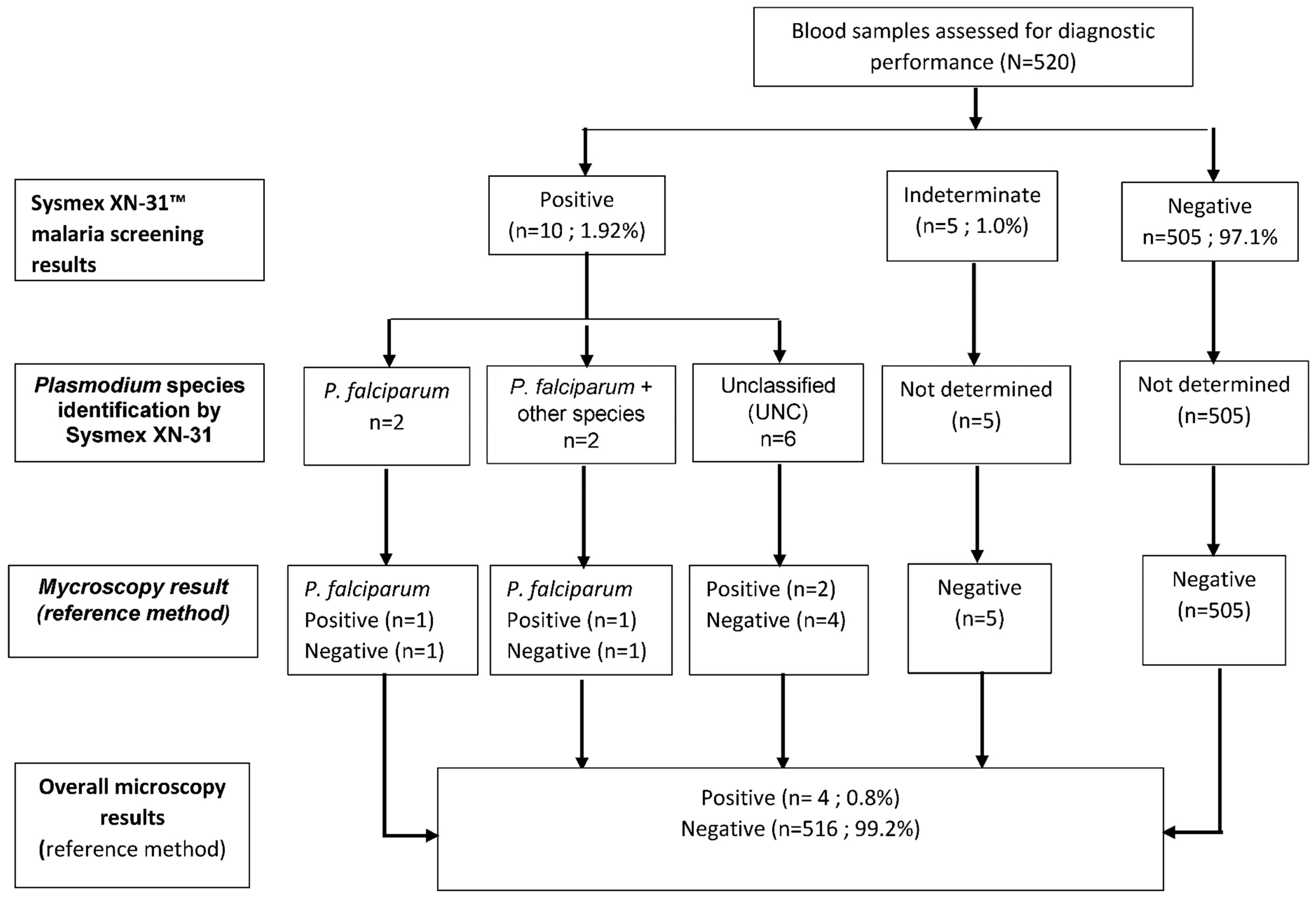
Malaria prevalence detected by Sysmex XN-31™ was 1.92% (10/520; 95% CI: 1.04–3.50) compared to 0.77% (4/520; 95% CI: 0.30–1.96) by microscopy. The median parasitemia measured by Sysmex XN-31™ was 98 parasites/µL (IQR: 37–229) compared to 140.5 parasites/µL (IQR: 31–250) by microscopy (Table 1).

Table 1.
Parasite density (median and IQR) for 10 cases detected by Sysmex XN-31™ and 4 cases by microscopy.
Table 1.
Parasite density (median and IQR) for 10 cases detected by Sysmex XN-31™ and 4 cases by microscopy.
| Method | N Positive | Median (Parasites/µL) | IQR (Parasites/µL) |
|---|---|---|---|
| Sysmex XN-31™ | 10 | 98 | 37–229 |
| Microscopy | 4 | 140.5 | 31–250 |
Among the ten Sysmex-positive cases, 5/10 (50%) had low parasitemia (20–100 parasites/µL), 4/10 (40%) had moderate parasitemia (100–1000 p/µL), and 1/10 (10%) had high parasitemia (>1000 parasites/µL). No sample with parasitemia below the analytical detection threshold (<20 parasites/µL) was detected by Sysmex XN-31™. Although one case showed nearly identical counts by both methods (415 parasites/µL by Sysmex XN-31™ compared to 422 parasites/µL by microscopy), the overall correlation analysis between Sysmex XN-31™ and microscopy parasitemias indicated a weak positive relationship that was not statistically significant (Spearman ρ = 0.25; p = 0.55).
Regarding species identification (Table 2), automated species identification was achieved in 4 out of 10 positive cases: two Plasmodium (P) falciparum monoinfections and two co-infections involving P. falciparum and at least one other species. Microscopy identified P. falciparum in the same two monoinfection cases but detected no co-infection cases. Additionally, 5 (1.0%) of the 520 samples yielded indeterminate Sysmex results (Table 2). None were confirmed positive by microscopy; they were excluded from the primary performance analysis to ensure robustness of the results.
Table 2.
Plasmodium species identification by Sysmex XN-31™ and by microscopy.
| Method | N Positive | P. falciparum Only | Co-Infections | Unclassified | Indeterminate Cases * |
|---|---|---|---|---|---|
| n (%) | n (%) | n (%) | n (%) | ||
| Sysmex XN-31™ | 10 | 2 (20) | 2 (20) | 6 (60) | 5 (1.0% of 520) |
| Microscopy | 4 | 4 (100) | 0 (0) | 0 (0) | 0 |
* Indeterminate cases: samples in which the Sysmex XN-31™ signal was considered non-interpretable.
When evaluating its performance against microscopy as the reference (Table 3), Sysmex XN-31™ achieved a sensitivity of 100% (95% CI: 39.8–100), a specificity of 98.8% (95% CI: 97.4–99.5), a positive predictive value (PPV) of 40% (95% CI: 12.2–73.8), and a negative predictive value (NPV) of 100% (95% CI: 99.3–100). Youden’s index was 0.99, the positive likelihood ratio (LR+) was 84.03, and the negative likelihood ratio (LR–) was 0.00.
Table 3.
Diagnostic performance indicators of Sysmex XN-31™ for the detection of asymptomatic malaria.
Table 3.
Diagnostic performance indicators of Sysmex XN-31™ for the detection of asymptomatic malaria.
| Indicator | Value | 95% Confidence Interval (Clopper–Pearson) |
|---|---|---|
| Sensitivity | 100.0% | [39.8–100%] |
| Specificity | 98.81% | [97.5–99.6%] |
| Positive predictive value (PPV) | 40.0% | [12.2–73.8%] |
| Negative predictive value (NPV) | 100.0% | [99.3–100%] |
| Positive likelihood ratio (LR+) | 84.03 | – |
| Negative likelihood ratio (LR−) | 0.00 | – |
| Youden’s index | 0.99 | – |
The agreement between the different diagnostic tests, estimated by Cohen’s kappa test, was κ = 0.57 (95% CI: 0.33–0.80). This indicates a moderate agreement between the two methods. Among the four microscopy-positive samples, one gametocyte carrier was noted (Table 4).
The case-by-case analysis (Table 4) showed that all microscopy-positive cases, including one gametocyte carrier, were among the ten Sysmex-positive cases; however, several low-parasitemia Sysmex-positive cases were negative via microscopy.
Table 4.
Results for blood of individual donors, tested by Sysmex XN-31™ and microscopy (N = 520).
| Donor ID | Sysmex XN-31 Result | MI-RBC% | MI-RBC# (P/µL) | Plasmodium Species Sysmex XN-31 | Microscopy Result (Thick Smear) | Parasitemia (P/µL) (Thick Smear) | Plasmodium Species (Thin Smear) | Parasite Stage (Thin Smear) |
|---|---|---|---|---|---|---|---|---|
| CRTS042 | Positive | 0.0011 | 56 | Unclassified | Negative | 0 | Negative | Negative |
| CRTS072 | Positive | 0.0018 | 98 | Unclassified | Negative | 0 | Negative | Negative |
| CRTS155 | Indeterminate | 0 | 0 | Not determined | Negative | 0 | Negative | Negative |
| CRTS157 | Indeterminate | 0 | 0 | Not determined | Negative | 0 | Negative | Negative |
| CRTS162 | Indeterminate | 0 | 0 | Not determined | Negative | 0 | Negative | Negative |
| CRTS194 | Positive | 0.0048 | 229 | P. falciparum | Positive | 31 | P. falciparum | Trophozoite |
| CRTS201 | Positive | 0.0463 | 2391 | P. falciparum + other species | Negative | 0 | Negative | Negative |
| CRTS203 | Positive | 0.0029 | 160 | P. falciparum | Negative | 0 | Negative | Negative |
| CRTS229 | Positive | 0.0008 | 29 | Unclassified | Negative | 0 | Negative | Negative |
| CRTS232 | Positive | 0.0100 | 415 | P. falciparum + other species | Positive | 422 | P. falciparum | Trophozoite |
| CRTS316 | Indeterminate | 0 | 0 | Not determined | Negative | 0 | Negative | Negative |
| CRTS320 | Positive | 0.0005 | 24 | Unclassified | Negative | 0 | Negative | Negative |
| CRTS402 | Positive | 0.0045 | 175 | Unclassified | Positive | ND | P. falciparum | Gametocytes |
| CRTS441 | Positive | 0.0008 | 37 | Unclassified | Negative | 250 | P. falciparum | Trophozoite |
| CRTS484 | Indeterminate | 0 | 0 | Not determined | Negative | 0 | Negative | Negative |
Caption. MI-RBC%: percentage of malaria-infected red blood cells, measured by Sysmex XN-31™. MI-RBC#: absolute number of malaria-infected red blood cells, expressed as parasites per microliter (P/μL), automatically estimated by Sysmex XN-31™. Sysmex XN-31™ results: Positive: parasitemia ≥ 20 infected red blood cells per microliter (iRBCs/μL), corresponding to the validated analytical threshold for malaria positivity; Indeterminate: cytometric profile not interpretable due to abnormal scattergram patterns or insufficient/ambiguous signal; these cases did not meet criteria for classification as positive or negative. Plasmodium species (Sysmex XN-31™): P. falciparum or P. falciparum + other species: automated identification provided by the analyzer; Unclassified (UNC): parasitemia ≥ 20 iRBCs/μL detected, but automated species identification not achieved; Not determined: species identification not possible due to indeterminate cytometric profile and/or absence of an interpretable parasitemia signal. Microscopy (thick smear): Result of thick-blood-smear examination (positive or negative). Parasitemia reported as parasites per microliter (Ps/μL) when positive. ND = not determined. Plasmodium species (thin smear): Species identified on thin-smear examination. “Negative” indicates that no malaria parasite was detected on microscopy and, consequently, no species could be identified. Parasite stage (thin smear): Developmental stage observed (e.g., trophozoite and gametocyte). When reported as “Negative”, this indicates that no parasite was detected on microscopy, and therefore no developmental stage could be identified. P/μL: Parasites per microliter.
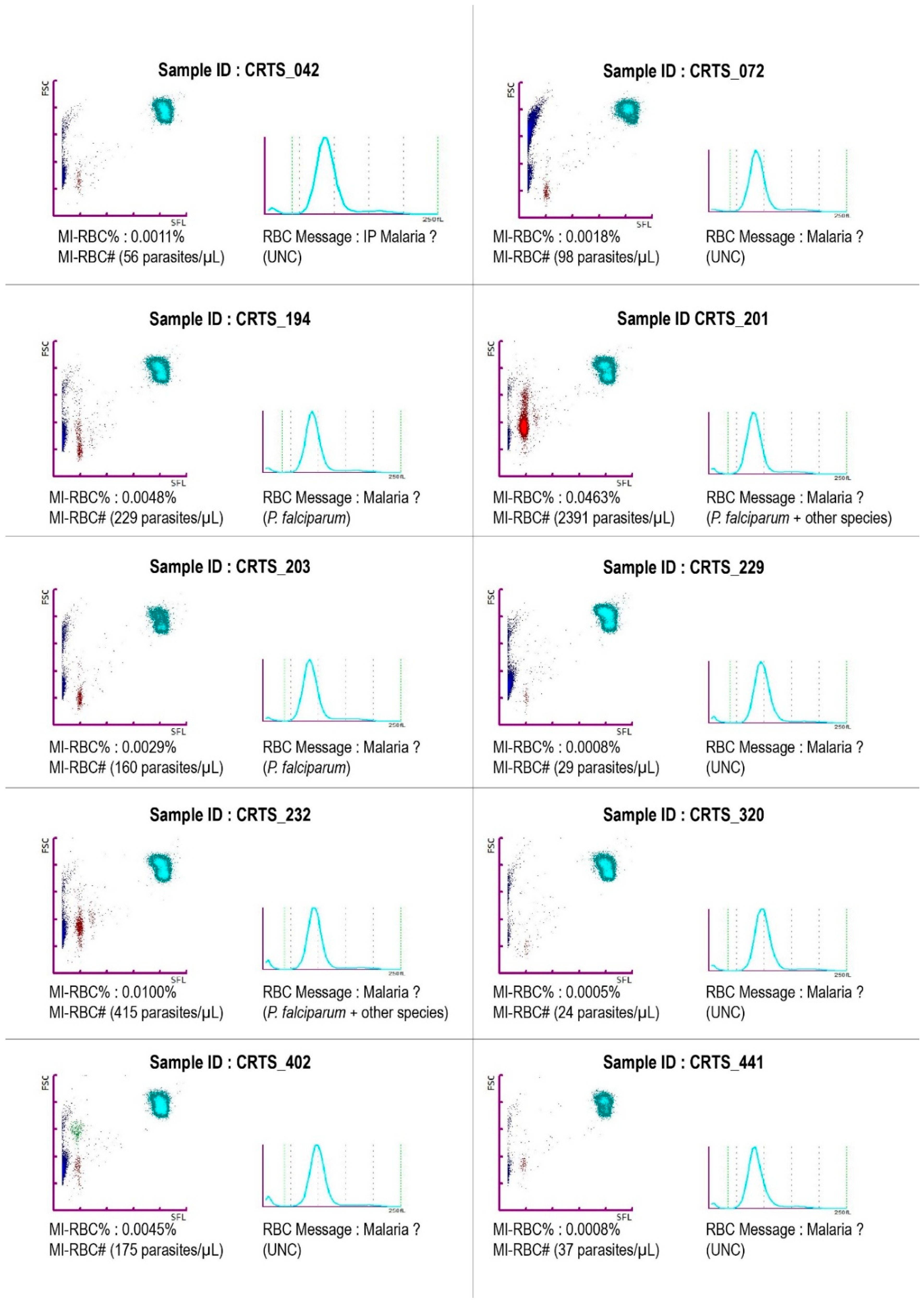
The cytometry scattergrams and corresponding parasite population ratios (MI-RBC%) for the 10 Sysmex XN-31™-positive samples are shown in Figure 2 to illustrate the analytical basis of automated detection.
Figure 2.
Representative cytometry profiles of the 10 Sysmex XN-31™-positive blood donor samples. Caption: Each panel displays the MI-RBC scattergram (upper left) and corresponding RBC histogram (upper right) obtained in low-malaria (LM) mode. The MI-RBC% (percentage of infected red blood cells) and MI-RBC# (absolute number of infected red blood cells expressed as parasites/µL) are indicated for each sample. Instrument malaria flags and classification messages are shown as reported by the analyzer (e.g., Malaria? (UNC), P. falciparum, or P. falciparum + other species). The panels illustrate the range of parasite densities detected in this cohort, from low-level parasitemia (<100 parasites/µL) to higher parasite burdens (>2000 parasites/µL). In the MI-RBC scattergram, red dots represent malaria-infected red blood cells (iRBCs), while blue dots correspond to non-infected red blood cells.
Figure 2.
Representative cytometry profiles of the 10 Sysmex XN-31™-positive blood donor samples. Caption: Each panel displays the MI-RBC scattergram (upper left) and corresponding RBC histogram (upper right) obtained in low-malaria (LM) mode. The MI-RBC% (percentage of infected red blood cells) and MI-RBC# (absolute number of infected red blood cells expressed as parasites/µL) are indicated for each sample. Instrument malaria flags and classification messages are shown as reported by the analyzer (e.g., Malaria? (UNC), P. falciparum, or P. falciparum + other species). The panels illustrate the range of parasite densities detected in this cohort, from low-level parasitemia (<100 parasites/µL) to higher parasite burdens (>2000 parasites/µL). In the MI-RBC scattergram, red dots represent malaria-infected red blood cells (iRBCs), while blue dots correspond to non-infected red blood cells.
4. Discussion
Our study demonstrates that Sysmex XN-31™ demonstrated an observed sensitivity of 100% in this dataset, although the precision of this estimate is limited by the small number of microscopy-confirmed positive cases. The analyzer also showed high specificity (98.8%) for detecting asymptomatic malaria among blood donors, with a reassuring NPV of 100% for transfusion safety. As expected in a low-prevalence setting, the PPV is low (40%). Sysmex XN-31™ demonstrated strong discriminative capacity, evidenced by a Youden’s index of 0.99 and a positive likelihood ratio of 84.03. The findings are consistent with three additional studies—one conducted in India [31] and two in Malawi—that indicated an automated detection rate that was double that of traditional microscopy [32,33]. A study in Southeast Asia shows sensitivity and specificity comparable to our findings regarding imported malaria [23]. In our study, 50% of Sysmex-positive samples had parasitemia below 100 parasites/µL—levels that are frequently undetectable by standard microscopy [32,33]. Sysmex XN-31TM’s ability to detect submicroscopic infections contributes to the identification of a latent reservoir of chronic carriers and may explain its apparent superior performance compared with microscopy. The absence of a significant correlation on parasitemias between Sysmex XN-31™ and microscopy (ρ = 0.25; p = 0.55) underscores the limitations of the microscopy method in detecting low parasite densities. Sysmex XN-31™ was able to type species in only 40% of positive cases, indicating its analytical thresholds at very low parasitemia [21]. Our findings confirm a decline in species-typing capacity at low densities [34] and reinforces the need for complementary methods such as thin-smear or molecular examination when precise species identification is required. This limitation highlights the complementary role of thin smears for reliable morphological identification and detection of transmissible stages [29,35]. Our investigation did not assess factors that can affect species-typing performance, such as sample quality, storage conditions, instrument calibration, or maintenance [21,25]. All indeterminate Sysmex XN-31™ results (1%) in our investigation were confirmed to be negative using microscopy, although the absence of molecular confirmation may have limited the detection of very-low-density infections. Molecular confirmation using PCR or LAMP, or expert slide review, would further strengthen discordant-case evaluation [36].
The inability of Sysmex XN-31™ to differentiate gametocytes from asexual stages represents an important limitation for accurately assessing post-transfusion transmission risk but does not affect its capacity to detect malaria-infected blood units [37]. Previous studies on Sysmex XN-31™ have focused on the detection and quantification of asexual Plasmodium stages, neglecting its efficacy in identifying or quantifying gametocytes [21,22]. The analyzer measures all infected red blood cells without differentiating stages, which is a recognized limitation [24].
Molecular techniques like quantitative reverse transcriptase–PCR (qRT-PCR) and quantitative nucleic acid sequence-based amplification (QT-NASBA), which target P. falciparum gametocyte-specific transcripts (Pfs25 mRNA and Pfs230p) and offer improved sensitivity and specificity, even at low densities, that enable accurate quantification of the infectious reservoir [38,39]. The techniques are costly, necessitate considerable technical expertise, and are infrequently accessible in transfusion centers in West Africa. The absence of a gametocyte-specific detection capability with Sysmex XN-31™ may limit the precise estimation of the infectious reservoir among asymptomatic donors. The integration of targeted gametocyte detection with molecular assays has the potential to enhance transfusion safety and support malaria elimination initiatives.
Limitations
Compared with studies based on smaller sample sizes and a limited number of malaria-positive cases, our study yielded wider confidence intervals for sensitivity estimates, reflecting lower statistical precision. This is a common limitation in low-prevalence settings such as blood donor populations. Seasonal variations in malaria transmission may also influence the number of asymptomatic infections detected, and future evaluations conducted at different periods of the year would provide a more comprehensive assessment of diagnostic performance [33]. A major methodological limitation of this study is the absence of molecular confirmation using a reference method such as polymerase chain reaction (PCR). Although expert microscopy was used as the comparator, it is well recognized that microscopy may fail to detect chronic low-density parasitemia, particularly among asymptomatic carriers. Consequently, it was not possible to reliably distinguish true Sysmex XN-31™ false-positive results from genuine submicroscopic infections detected only by the analyzer. At the time of the study, no frozen aliquots suitable for retrospective PCR analysis had been prospectively stored, which precluded molecular confirmation of Sysmex-positive samples. Conversely, without external molecular validation, rare analytical artifacts or false-positive signals generated by automated hematology analyzers cannot be definitively excluded. Several authors have therefore recommended the systematic inclusion of molecular reference methods in evaluations of new malaria diagnostic technologies [32,33,38].
In addition, parasitemia estimation based solely on thick-smear microscopy may introduce measurement variability related to operator experience and the volume of blood examined, particularly at low parasite densities [40]. Thin-smear examination was primarily used for species identification and stage confirmation and is less reliable for quantitative assessment. Finally, the generalizability of our findings remains limited because this study did not include a cost-effectiveness analysis, an operational feasibility assessment, or an evaluation of the potential impact on blood transfusion safety workflows. Future research should therefore integrate molecular confirmation methods, preserve aliquots for retrospective testing, and assess the financial, logistical, and organizational implications of implementing Sysmex XN-31™ in transfusion settings.
Assessing parasitemia solely with the thick-smear method may introduce bias due to operator-related variability and the volume of blood tested, particularly at low densities [40]. The thin smear is mainly used for morphological identification and stage confirmation and is therefore less robust for quantitative analysis at low parasitemia.
Moreover, our findings are not broadly applicable or generalizable due to the lack of a cost-effective analysis, an organizational assessment, and a study of the impact of the transfusion process. Future research should examine the financial, logistical, and organizational implications of relying on Sysmex XN-31TM in blood transfusion settings, in addition to evaluating its analytical capabilities.
5. Conclusion
Sysmex XN-31™ exhibited a sensitivity of 100% and a specificity of 98.8% in the detection of asymptomatic malaria in blood donors, even at low levels of parasitemia. It is a rapid and automated tool that shows potential for improving transfusion safety in endemic regions. Large-scale implementation requires: molecular validation of positive and indeterminate results, a cost-effectiveness analysis adapted to local resources, and focused training for users. Multicenter studies carried out particularly during high-transmission seasons and incorporating economic evaluations are critical for the broad adoption and scalability of the standardized screening protocol.
Abbreviations
EDTA: ethylenediaminetetraacetic acid; IQR: interquartile range; iRBC: infected red blood cell; LAMP: loop-mediated isothermal amplification; LR–: negative likelihood ratio; LR+: positive likelihood ratio; MI-RBCs: malaria-infected red blood cells; NPV: negative predictive value; PCR: polymerase chain reaction; Pfs230p: Plasmodium falciparum gametocyte surface protein 230, precursor; Pfs25 mRNA: messenger RNA for Plasmodium falciparum surface protein 25; PPV: positive predictive value; qPCR: quantitative polymerase chain reaction; QT-NASBA: quantitative nucleic acid sequence-based amplification; RBTC: Regional Blood Transfusion Center; RDT: rapid diagnostic test; qRT-PCR: quantitative reverse transcriptase–polymerase chain reaction.
Author Contributions
P.C.K.B. and E.Y. conceived and planned the study and conducted the literature review. E.Y. and L.H.E.K. collected field data and performed laboratory analyses under the supervision of P.C.K.B. P.C.K.B. analyzed the data and drafted the first version of the manuscript. K.E.A., A.J.S.M., V.A.B.-T., G.M.E.K., A.H.V.-B., F.K.K., A.K.-T., V.D., E.I.H.M. and W.Y. critically revised the manuscript. All authors have read and agreed to the published version of the manuscript.
Funding
The Sysmex XN-31™ analyzer used in this study was part of the routine laboratory equipment of the participating institution. No financial support, reagents, or technical assistance were provided by the manufacturer.
Acknowledgments
We thank the team at the Regional Blood Transfusion Center (Abidjan) for their invaluable support in recruiting study participants and for providing access to Sysmex XN-31 for the screening of asymptomatic malaria carriage among blood donors.
Conflicts of Interest
The authors declare that Sysmex Corporation had no role in the study design, data collection, data analysis, interpretation of results, or manuscript preparation.
References
- Direction de la Météorologie Nationale. Bulletin de Veille Climatique En Côte d’Ivoire: Mois D’octobre 2024; N° 10—Année 2024; SODEXAM: Abidjan, Côte d’Ivoire, 2024; p. 18. [Google Scholar]
- Doumbia, M.; Coulibaly, J.T.; Silué, D.K.; Cissé, G.; N’Dione, J.-A.; Koné, B. Effects of Climate Variability on Malaria Transmission in Southern Côte d’Ivoire, West Africa. Int. J. Environ. Res. Public Health 2023, 20, 7102. [Google Scholar] [CrossRef] [PubMed]
- Gouamene, T.J.R.; Edi, C.A.V.; Kwadio, E.; Gbalegba, C.G.N.; Zahouli, J.Z.B.; Koudou, B.G. Malaria Transmission in the Coastal Zone and in the Centre of Côte d’Ivoire during the Dry Season. Malar. J. 2024, 23, 400. [Google Scholar] [CrossRef] [PubMed]
- World Health Organization. Malaria 2024 Côte d’Ivoire Country Profile; Technical Document; World Health Organization: Geneva, Switzerland, 2024; Available online: https://www.who.int/publications/m/item/malaria-2024-civ-country-profile (accessed on 22 April 2026).
- Lindblade, K.A.; Steinhardt, L.; Samuels, A.; Kachur, S.P.; Slutsker, L. The Silent Threat: Asymptomatic Parasitemia and Malaria Transmission. Expert Rev. Anti-Infect. Ther. 2013, 11, 623–639. [Google Scholar] [CrossRef] [PubMed]
- White, N.J.; Pukrittayakamee, S.; Hien, T.T.; Faiz, M.A.; Mokuolu, O.A.; Dondorp, A.M. Malaria. Lancet 2014, 383, 723–735. [Google Scholar] [CrossRef] [PubMed]
- Whittaker, C.; Slater, H.; Nash, R.; Bousema, T.; Drakeley, C.; Ghani, A.C.; Okell, L.C. Global Patterns of Submicroscopic Plasmodium falciparum Malaria Infection: Insights from a Systematic Review and Meta-Analysis of Population Surveys. Lancet Microbe 2021, 2, e366–e374. [Google Scholar] [CrossRef] [PubMed]
- Sagna, A.B.; Gebre, Y.; Vera-Arias, C.A.; Traoré, D.F.; Tchekoi, B.N.; Assi, S.B.; Koffi, A.A.; Rogier, C.; Remoue, F.; Koepfli, C. High Prevalence of Asymptomatic and Subpatent Plasmodium falciparum Infections but No Histidine-Rich Protein 2 Gene Deletion in Bouaké, Côte d’Ivoire. Sci. Rep. 2024, 14, 19060. [Google Scholar] [CrossRef] [PubMed]
- Gbalégba, C.G.N.; Ba, H.; Silué, K.D.; Ba, O.; Tia, E.; Chouaibou, M.; Tian-Bi, N.T.Y.; Yapi, G.Y.; Koné, B.; Utzinger, J.; et al. Distribution of Plasmodium spp. Infection in Asymptomatic Carriers in Perennial and Low Seasonal Malaria Transmission Settings in West Africa. Infect. Dis. Poverty 2018, 7, 39. [Google Scholar] [CrossRef] [PubMed]
- Shankar, H.; Singh, M.P.; Phookan, S.; Singh, K.; Mishra, N. Diagnostic Performance of Rapid Diagnostic Test, Light Microscopy and Polymerase Chain Reaction during Mass Survey Conducted in Low and High Malaria-Endemic Areas from Two North-Eastern States of India. Parasitol. Res. 2021, 120, 2251–2261. [Google Scholar] [CrossRef] [PubMed]
- Naing, C.; Htet, N.H.; Aye, S.N.; Aung, H.H.; Tanner, M.; Whittaker, M.A. Detection of Asymptomatic Malaria in Asian Countries: A Meta-Analysis of Diagnostic Accuracy. Malar. J. 2022, 21, 50. [Google Scholar] [CrossRef] [PubMed]
- Uneke, C.; Ogbu, O.; Nwojiji, V. Potential Risk of Induced Malaria by Blood Transfusion in South-Eastern Nigeria. McGill J. Med. 2020, 9, 8–13. [Google Scholar] [CrossRef]
- Nansseu, J.R.N.; Noubiap, J.J.N.; Ndoula, S.T.; Zeh, A.F.M.; Monamele, C.G. What Is the Best Strategy for the Prevention of Transfusion-Transmitted Malaria in Sub-Saharan African Countries Where Malaria Is Endemic? Malar. J. 2013, 12, 465. [Google Scholar] [CrossRef] [PubMed]
- Owusu-Ofori, A.K.; Betson, M.; Parry, C.M.; Stothard, J.R.; Bates, I. Transfusion-Transmitted Malaria in Ghana. Clin. Infect. Dis. 2013, 56, 1735–1741. [Google Scholar] [CrossRef] [PubMed]
- Dou, G.; Aka, N.; Adjo, G.; Nassoue, O.; Tamboura, M.; N’guessan, A.; Anzara, A.; Kouakou, A.; Zika, K.; Adoubryn, K. Asymptomatic Carriage of Plasmodium spp. and Blood Microfilariae among Blood Donors at the National Blood Transfusion Center (NBTC) in Abidjan in 2022. ASEAN J. Psychiatry 2024, 01–06. [Google Scholar] [CrossRef]
- Opoku Afriyie, S.; Addison, T.K.; Gebre, Y.; Mutala, A.-H.; Antwi, K.B.; Abbas, D.A.; Addo, K.A.; Tweneboah, A.; Ayisi-Boateng, N.K.; Koepfli, C.; et al. Accuracy of Diagnosis among Clinical Malaria Patients: Comparing Microscopy, RDT and a Highly Sensitive Quantitative PCR Looking at the Implications for Submicroscopic Infections. Malar. J. 2023, 22, 76. [Google Scholar] [CrossRef] [PubMed]
- Zhang, J.; Chen, X.; Pan, M.; Qin, Y.; Zhao, H.; Yang, Q.; Li, X.; Zeng, W.; Xiang, Z.; Wu, Y.; et al. Application of a Low-Cost, Specific, and Sensitive Loop-Mediated Isothermal Amplification (LAMP) Assay to Detect Plasmodium falciparum Imported from Africa. Mol. Biochem. Parasitol. 2022, 252, 111529. [Google Scholar] [CrossRef] [PubMed]
- Kamau, E.; Tolbert, L.S.; Kortepeter, L.; Pratt, M.; Nyakoe, N.; Muringo, L.; Ogutu, B.; Waitumbi, J.N.; Ockenhouse, C.F. Development of a Highly Sensitive Genus-Specific Quantitative Reverse Transcriptase Real-Time PCR Assay for Detection and Quantitation of Plasmodium by Amplifying RNA and DNA of the 18S rRNA Genes. J. Clin. Microbiol. 2011, 2946–2953. [Google Scholar] [CrossRef] [PubMed]
- Yalley, A.K.; Ocran, J.; Cobbinah, J.E.; Obodai, E.; Yankson, I.K.; Kafintu-Kwashie, A.A.; Amegatcher, G.; Anim-Baidoo, I.; Nii-Trebi, N.I.; Prah, D.A. Advances in Malaria Diagnostic Methods in Resource-Limited Settings: A Systematic Review. Trop. Med. Infect. Dis. 2024, 9, 190. [Google Scholar] [CrossRef] [PubMed]
- Hopkins, H.; González, I.J.; Polley, S.D.; Angutoko, P.; Ategeka, J.; Asiimwe, C.; Agaba, B.; Kyabayinze, D.J.; Sutherland, C.J.; Perkins, M.D.; et al. Highly Sensitive Detection of Malaria Parasitemia in a Malaria-Endemic Setting: Performance of a New Loop-Mediated Isothermal Amplification Kit in a Remote Clinic in Uganda. J. Infect. Dis. 2013, 208, 645–652. [Google Scholar] [CrossRef] [PubMed]
- Khartabil, T.A.; de Rijke, Y.B.; Koelewijn, R.; van Hellemond, J.J.; Russcher, H. Fast Detection and Quantification of Plasmodium Species Infected Erythrocytes in a Non-Endemic Region by Using the Sysmex XN-31 Analyzer. Malar. J. 2022, 21, 119. [Google Scholar] [CrossRef] [PubMed]
- Komaki-Yasuda, K.; Kutsuna, S.; Kawaguchi, M.; Kamei, M.; Uchihashi, K.; Nakamura, K.; Nakamoto, T.; Ohmagari, N.; Kano, S. Clinical Performance Testing of the Automated Haematology Analyzer XN-31 Prototype Using Whole Blood Samples from Patients with Imported Malaria in Japan. Malar. J. 2022, 21, 229. [Google Scholar] [CrossRef] [PubMed]
- Picot, S.; Perpoint, T.; Chidiac, C.; Sigal, A.; Javouhey, E.; Gillet, Y.; Jacquin, L.; Douplat, M.; Tazarourte, K.; Argaud, L.; et al. Diagnostic Accuracy of Fluorescence Flow-Cytometry Technology Using Sysmex XN-31 for Imported Malaria in a Non-Endemic Setting. Parasite 2022, 29, 31. [Google Scholar] [CrossRef] [PubMed]
- Dumas, C.; Tirard-Collet, P.; Mestrallet, F.; Girard, S.; Jallades, L.; Picot, S.; Bienvenu, A.-L. Flagging Performance of Sysmex XN-10 Haematology Analyser for Malaria Detection. J. Clin. Pathol. 2020, 73, 676–677. [Google Scholar] [CrossRef] [PubMed]
- Zuluaga-Idárraga, L.; Rios, A.; Sierra-Cifuentes, V.; Garzón, E.; Tobón-Castaño, A.; Takehara, I.; Toya, Y.; Izuka, M.; Uchihashi, K.; Lopera-Mesa, T.M. Performance of the Hematology Analyzer XN-31 Prototype in the Detection of Plasmodium Infections in an Endemic Region of Colombia. Sci. Rep. 2021, 11, 5268. [Google Scholar] [CrossRef] [PubMed]
- Sawadogo, S.; Hwandih, T.; Saker, J.; Coetzer, T.L.; Paszkowska, A.; Kompaoré, S.C.B.; Münster, M. Blood Donors as a Sentinel Population for Real-Time Malaria Surveillance Using the Sysmex XN-31: A One-Year Review from the National Blood Transfusion Centre in Burkina Faso. Malar. J. 2025, 24, 288. [Google Scholar] [CrossRef] [PubMed]
- Tokponnon, T.F.; Razaki, O.; Denon, Y.E.; Yerima, I.J.G.; Festus, H.; Brunelle, A.; Thibaud, L.; Kpemasse, A.; Dagbadji, M.F.; Padonou, G.G.; et al. Evaluation of the Diagnostic Capacity of Malaria by XN-31 Automate at Cotonou in an Endemic Area of Benin, West Africa. Adv. Infect. Dis. 2024, 14, 810–821. [Google Scholar] [CrossRef]
- Buderer, N.M. Statistical Methodology: I. Incorporating the Prevalence of Disease into the Sample Size Calculation for Sensitivity and Specificity. Acad. Emerg. Med. 1996, 895–900. [Google Scholar] [CrossRef] [PubMed]
- World Health Organization. Microscopie Pour La Détection, l’identification et La Quantification Des Parasites Du Paludisme Sur Frottis Sanguins Ou Gouttes Épaisses Colorés Dans Les Environnements de Recherche: Procédures: Guide Méthodologique; World Health Organization: Geneva, Switzerland, 2020; Available online: https://iris.who.int/handle/10665/331144 (accessed on 22 April 2026).
- Landis, J.R.; Koch, G.G. The Measurement of Observer Agreement for Categorical Data. Biometrics 1977, 33, 159–174. [Google Scholar] [CrossRef]
- Tiwari, A.K.; Goel, S.; Singh, G.; Gahlot, P.K.; Saxena, R.; Jadhav, V.; Sethi, M. Evaluation of New Haematology Analyser, XN-31, for Malaria Detection in Blood Donors: A Single-Centre Study from India. Vox Sang. 2024, 119, 556–562. [Google Scholar] [CrossRef] [PubMed]
- M’baya, B.; Mfune, T.; Samon, A.; Hwandih, T.; Münster, M. Evaluation of the Sysmex XN-31 Automated Analyser for Blood Donor Malaria Screening at Malawi Blood Transfusion Services. Vox Sang. 2022, 117, 346–353. [Google Scholar] [CrossRef] [PubMed]
- Kayange, M.; M’baya, B.; Hwandih, T.; Saker, J.; Coetzer, T.L.; Münster, M. Automated Measurement of Malaria Parasitaemia among Asymptomatic Blood Donors in Malawi Using the Sysmex XN-31 Analyser: Could Such Data Be Used to Complement National Malaria Surveillance in Real Time? Malar. J. 2022, 21, 299. [Google Scholar] [CrossRef] [PubMed]
- Kabalu Tshiongo, J.; Luzolo, F.; Kabena, M.; Kuseke, L.; Djimde, M.; Mitashi, P.; Lumbala, C.; Kayentao, K.; Menting, S.; Mens, P.F.; et al. Performance of Ultra-Sensitive Malaria Rapid Diagnostic Test to Detect Plasmodium falciparum Infection in Pregnant Women in Kinshasa, the Democratic Republic of the Congo. Malar. J. 2023, 22, 322. [Google Scholar] [CrossRef] [PubMed]
- Centers for Disease Control and Prevention (CDC). Malaria Diagnostic Tests; CDC. Available online: https://www.cdc.gov/malaria/hcp/diagnosis-testing/malaria-diagnostic-tests.html (accessed on 22 April 2026).
- Tougan, T.; Suzuki, Y.; Itagaki, S.; Izuka, M.; Toya, Y.; Uchihashi, K.; Horii, T. An Automated Haematology Analyzer XN-30 Distinguishes Developmental Stages of Falciparum Malaria Parasite Cultured in Vitro. Malar. J. 2018, 17, 59. [Google Scholar] [CrossRef] [PubMed]
- Bousema, T.; Drakeley, C. Epidemiology and Infectivity of Plasmodium falciparum and Plasmodium Vivax Gametocytes in Relation to Malaria Control and Elimination. Clin. Microbiol. Rev. 2011, 24, 377–410. [Google Scholar] [CrossRef] [PubMed]
- Meerstein-Kessel, L.; Andolina, C.; Carrio, E.; Mahamar, A.; Sawa, P.; Diawara, H.; van de Vegte-Bolmer, M.; Stone, W.; Collins, K.A.; Schneider, P.; et al. A Multiplex Assay for the Sensitive Detection and Quantification of Male and Female Plasmodium falciparum Gametocytes. Malar. J. 2018, 17, 441. [Google Scholar] [CrossRef] [PubMed]
- Santolamazza, F.; Avellino, P.; Siciliano, G.; Yao, F.A.; Lombardo, F.; Ouédraogo, J.B.; Modiano, D.; Alano, P.; Mangano, V.D. Detection of Plasmodium falciparum Male and Female Gametocytes and Determination of Parasite Sex Ratio in Human Endemic Populations by Novel, Cheap and Robust RTqPCR Assays. Malar. J. 2017, 16, 468. [Google Scholar] [CrossRef] [PubMed]
- Dumas, C.; Bienvenu, A.-L.; Girard, S.; Picot, S.; Debize, G.; Durand, B. Automated Plasmodium Detection by the Sysmex XN Hematology Analyzer. J. Clin. Pathol. 2018, 71, 594–599. [Google Scholar] [CrossRef] [PubMed]
© 2026 Copyright by Authors. Licensed as an open access article using a CC BY 4.0 license.
